What Is Retinal Imaging and How Does It Work?
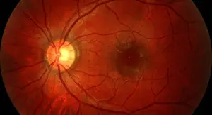
Retinal imaging is a non-invasive diagnostic procedure that uses specialized digital cameras and scanning technology to capture detailed images of the structures at the back of the eye. These structures include the retina itself (the light-sensitive tissue layer), the optic nerve head, the macula (responsible for central vision), and the blood vessels that supply the entire retinal network.
Modern retinal cameras use a combination of high-powered lenses, precise lighting, and digital sensors to photograph the fundus — the interior surface of the eye. Some systems, such as optical coherence tomography (OCT), go even further by creating a three-dimensional cross-sectional map of retinal layers at a microscopic level, revealing structural changes invisible to standard examination. Ultra-widefield imaging systems can capture up to 200 degrees of the retina in a single image, giving doctors a panoramic view they have never had before.
The procedure itself is remarkably simple and comfortable. Most retinal imaging is performed without eye drops or dilation, takes less than five minutes from start to finish, and involves nothing more than resting your chin on a support and looking at a small target light while the camera does its work. There is no pain, no pressure, and no recovery time. The images are instantly available on-screen for your doctor to review with you in real time.
The Retina: A Window Into Your Whole Body's Health
What makes the retina truly extraordinary is that it is the only place in the human body where blood vessels and nerve tissue can be observed directly, without surgery or invasive procedures. Because the retina shares circulatory and neurological characteristics with organs throughout the body, changes visible in the retinal vasculature can reflect diseases developing elsewhere — often years before other symptoms appear.
At Envision Eye Care, our eye doctors routinely identify early signs of systemic conditions through retinal imaging — conditions patients had no idea were developing. This window to your health makes retinal imaging one of the most powerful preventive health tools available today, not just an eye exam add-on.
Eye Diseases Detected Early Through Retinal Imaging
Managing eye disease starts with early detection — and retinal imaging is one of the most powerful tools we have to catch problems before they cause permanent damage.
Glaucoma is often called the silent thief of sight because it causes irreversible vision loss with virtually no warning symptoms until damage is advanced. Retinal imaging allows us to examine the optic nerve head in extraordinary detail, identifying characteristic changes in the cup-to-disc ratio, nerve fiber layer thinning, and optic disc hemorrhages that signal glaucomatous damage years before significant vision loss occurs. Early detection means early treatment — and treatment started early can preserve vision for a lifetime.
Age-Related Macular Degeneration (AMD) is the leading cause of irreversible central vision loss in adults over 50. The macula — the small, high-acuity region at the center of the retina — is vulnerable to the formation of drusen (protein deposits), abnormal blood vessel growth, and geographic atrophy. Retinal imaging pinpoints the earliest drusen formations and structural changes in the macula, allowing patients and doctors to intervene with lifestyle modifications, nutritional supplementation, and in the case of wet AMD, timely anti-VEGF injections that can halt or reverse vision loss.
Diabetic Retinopathy is the most common complication of diabetes and a leading cause of blindness in working-age adults. High blood sugar damages the tiny blood vessels of the retina, causing them to leak, swell, or grow abnormally. Retinal imaging can detect the very first microaneurysms and hemorrhages — signs of non-proliferative diabetic retinopathy — before you notice any change in your vision. For patients with diabetes, annual retinal imaging is not optional; it is essential. Caught early, diabetic retinopathy is highly treatable with laser therapy, injectable medications, and improved blood sugar management.
Hypertensive Retinopathy is the retinal damage caused by chronic high blood pressure. Elevated blood pressure strains and narrows the retinal blood vessels over time, causing changes visible in retinal photographs — arteriovenous nicking, copper or silver wiring of the arteries, flame hemorrhages, and cotton wool spots. These findings not only tell us your retinas are under stress but also signal that your cardiovascular system may need urgent attention.
Retinal Detachment and Tears can occur when the vitreous pulls away from the retinal surface, creating tears through which fluid seeps, causing the retina to lift from its underlying support tissue. If untreated, a full retinal detachment leads to profound and permanent vision loss. Widefield retinal imaging captures the peripheral retina where tears and detachments often begin — a region difficult or impossible to view with standard examination methods alone.
Why Early Detection Is a Game-Changer
The most important thing to understand about eye disease is that most causes of vision loss are preventable — but only when detected in time. The tragedy of conditions like glaucoma, macular degeneration, and diabetic retinopathy is not that they cannot be treated, but that they so often go undetected until substantial, irreversible damage has already occurred.
Studies consistently show that early-stage treatment of wet AMD reduces the risk of severe vision loss by more than 90%. Early glaucoma treatment can cut the rate of visual field loss by over 50%. For diabetic retinopathy, early laser treatment reduces the risk of blindness by up to 90%. These are not incremental improvements — these are the differences between a life with healthy vision and a life of significant visual impairment.
Retinal Imaging Is for Everyone — At Every Age
A common misconception is that retinal imaging is only relevant for older adults or patients with existing health conditions. In reality, retinal imaging provides valuable baseline and monitoring data for patients of all ages — including children and young adults.
Young patients can develop conditions such as juvenile-onset macular dystrophies, myopia-related retinal changes, and even early signs of systemic disease. Establishing a retinal baseline image in childhood or young adulthood creates a reference point that allows subtle future changes to be detected with far greater precision.
For adults with diabetes, hypertension, a family history of glaucoma or macular degeneration, high myopia, or those over age 40, annual retinal imaging is strongly recommended. For all other adults, retinal imaging every one to two years alongside a comprehensive eye exam provides comprehensive, proactive protection.
Make Retinal Imaging a Part of Your Routine Eye Care
A comprehensive eye exam that includes retinal imaging is one of the most valuable health screenings available — and one of the most underutilized. In the time it takes to complete your exam at Envision Eye Care, our advanced imaging technology creates a detailed, permanent digital record of your retinal health that we track and compare at every future visit. Changes are flagged early. Diseases are caught before they become crises.
The team at Envision Eye Care is committed to delivering the highest standard of preventive eye care to every patient — from children coming in for their first exam to adults managing chronic conditions. Our state-of-the-art retinal imaging technology, combined with the expertise of our doctors, gives you a level of insight into your eye health that simply is not possible with a traditional eye exam alone.
Do not wait for symptoms to tell you something is wrong. By then, the damage may already be done. Contact Envision Eye Care today to schedule your comprehensive eye exam with retinal imaging. Your vision is irreplaceable — let us help you protect it for life.